Making the Worlds of Wonder Data FAIR

The crowdsourced Worlds of Wonder data have been published open access on DataverseNL, an online data repository used by Dutch universities. In preparing the data for publication, the Worlds of Wonder researchers received help from the data stewards at Maastricht University, as well as the Open Science Community Maastricht, to make sure that the data would comply with the FAIR guidelines for data reuse. In the interview below, one of the researchers, Lea Beiermann, talks about her experience of making data FAIR.

The interview originally appeared as part of a series on FAIR data in FASoS Weekly, the newsletter of the Faculty of Arts and Social Sciences, Maastricht University (UM).

How did you find out about FAIR?

The first time I heard about FAIR was in a presentation by Michel Dumontier (at UM’s Institute of Data Science, IDS). After that, I saw that the UM library actively promoted the FAIR principles on social media. I also knew about FAIR because it was an important element of the data management plan that I had to write to receive funding for my PhD research.

What does FAIR mean to you?

From the data science perspective, FAIR describes data that are findable, accessible, interoperable, and reusable. As stated on the IDS website, FAIR is a good foundation to make data science more responsible. Ideally, it allows data to be shared widely.

For me, however, FAIR also means responsible data collection practices. During my PhD research, I noticed that this is an additional challenge, and that ethically sound data collection is sometimes not easily compatible with FAIR principles. I ran a web-based citizen science project as part of my PhD, which asked citizen scientists to help me analyse historical sources. According to the FAIR principles, I should have made sure that the crowdsourced data is reusable, and to some extent I did manage to do that, but sometimes it was difficult to turn the historical research of my citizen scientists into machine-readable data.

From a data scientist’s point of view, I probably would have got the most (re)usable clean data by having citizen scientists make yes/no classifications and tick boxes – quite monotonous tasks. A large part of my project consisted of such classification tasks, but I think the more interesting research (from a historian’s point of view, and also more interesting for the citizen scientists) happened in discussions in our chat forum. Of course, I can still make these discussions findable and accessible, for example by storing them with appropriate metadata, but they cannot be reused as easily as the more simple, machine-readable data.

Why did you decide to make your data FAIR?

Fair use of data is widely considered the default for citizen science projects. The consensus is that data that are crowdsourced should not belong to the principal researchers only but to everyone who helped compile them or who would like to reuse them. I reused the data collected through another citizen science project “Science Gossip” and found them a useful addition to my own data. I wanted to make it possible for others to reuse our project data too, so that is mainly why I decided to make my data FAIR.

How has the data steward (Maria Vivas Romero) helped you make your data FAIR?

Maria helped me understand and fill out UM’s data management plan – and I learned much more about FAIR data when we filled out the plan. Maria reminded me of the important role of metadata, such as commonly used keywords in my field of research, and that I could even make data findable whose use is restricted by copyright. For example, I could not share photos of archival material, but I could add metadata on these sources to my project to let other researchers know that they exist.

Was it a lot of work to make your data FAIR?

I am actually still in the process of making my data FAIR, even though I started my PhD a while ago. It does take quite some time, but for me this is absolutely worth it. After all, I used data collected by others, so it seems only fair to try and make my data as accessible and reusable as I could. And Maria has been a great help in making that possible.

How do you think making data FAIR benefits you?

I think there are several benefits to making your data FAIR. First, besides that I think it is only fair to make data available to others because I also make use of other researchers’ data, it also make my research and me as a researcher more findable and visible. Second, making data FAIR means that other research projects can benefit from all the work citizen scientists put into my project, so it gives greater value to their (and my) work. Third, FAIR data reuse can generate new research ideas that I did not think of but that might be interesting for my work.

How do you think making data FAIR helps other researchers?

Other researchers may reuse the historical data we collected, as well as the Python scripts one of my citizen scientists, Peter Mason, wrote to analyse the data. This means that others cannot only reuse historical data but also, possibly, learn from the way we analysed our data and do it in a similar way, or better. They can also get a clearer idea of the kind of data citizen science projects yield. I expected to get much cleaner data than we actually gathered – seeing the data I work with now in advance would have helped me to have more realistic expectations.

What hurdles did you come across when trying to make data FAIR?

I think making data FAIR does require quite some time. What I personally also experienced as a hurdle, or rather time-consuming, was combining the richness of qualitative historical research with machine-readability, or simple keywords. Moreover, I know that many of my colleagues collect sensitive data that they cannot share. On the other hand, we do have an excellent data steward who is happy to help us overcome these hurdles!
Meet the Illustrators

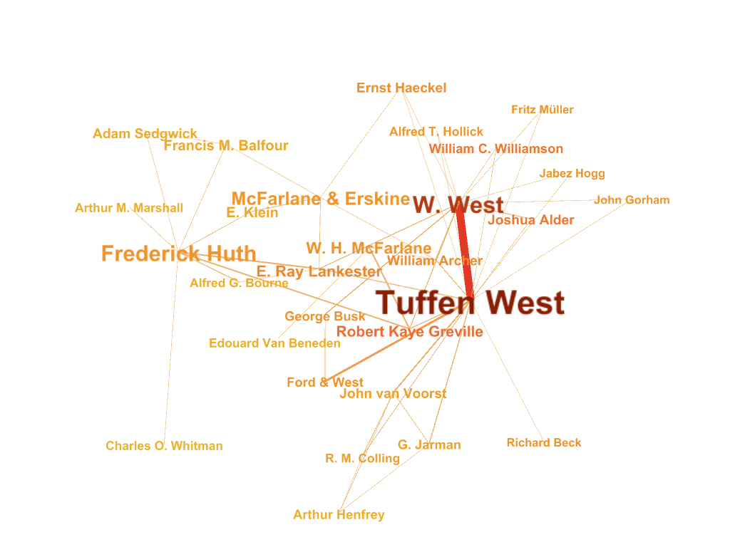
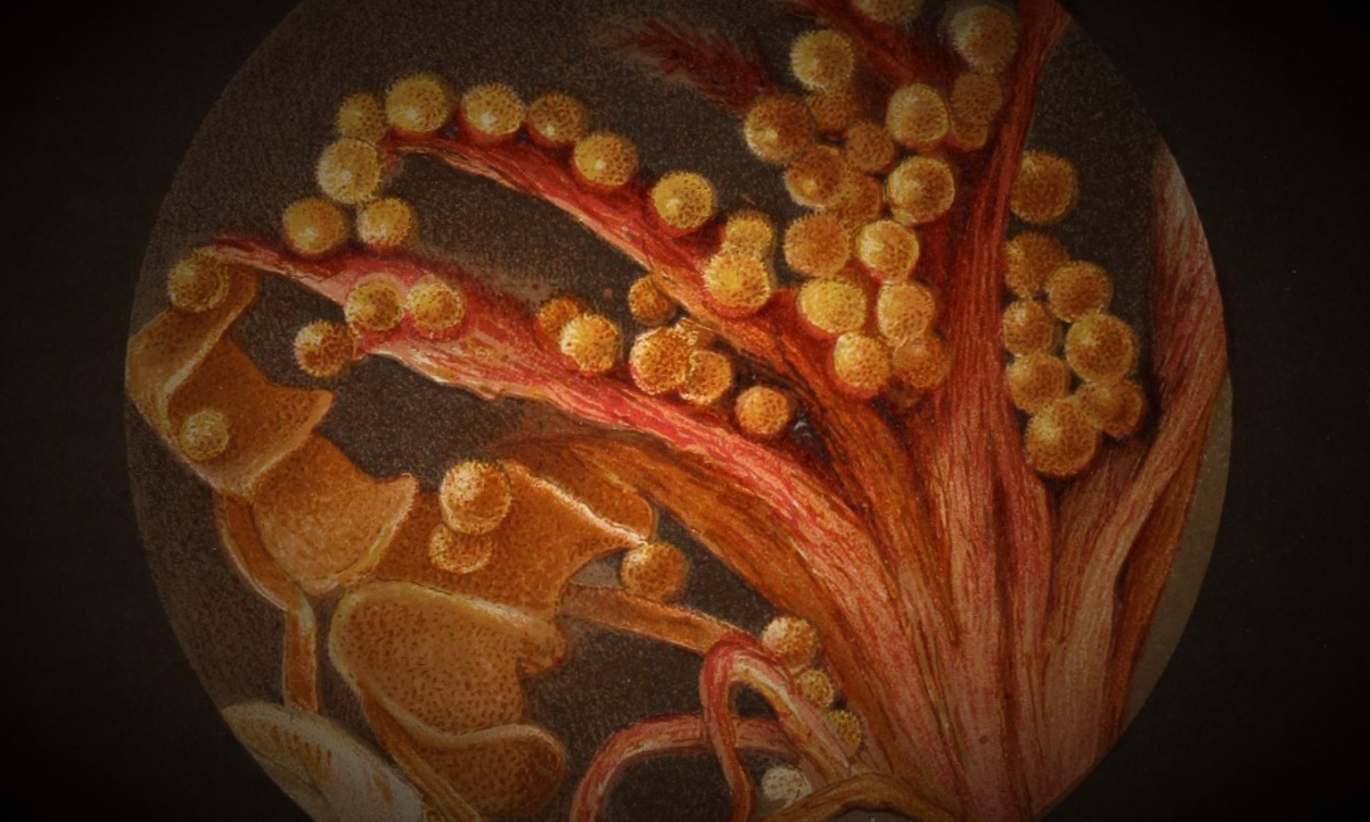
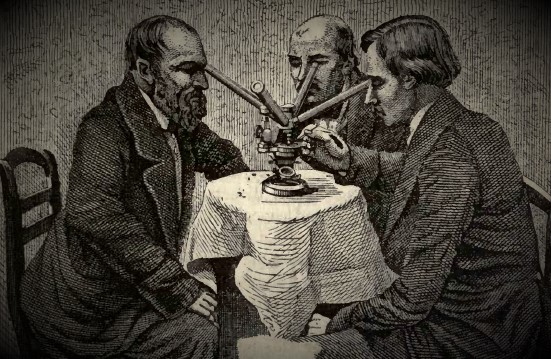
While you were busy collecting data in the Zooniverse, we started to aggregate and clean the data, so it could eventually be used to visualise the number and type of illustrations in a publication, as well as networks of illustrators. We were lucky to get a lot of help from Pmason, who wrote numerous Python scripts to analyse our data, and we thank him for that. Now, it’s time to finally meet the illustrators, engravers and printers of the illustrations you’ve been looking at!

The network above this text draws on the data you collected. You deciphered and transcribed the handwritten texts scribbled above or below illustrations, which often contained the names of the people who made the illustration. The network shows the most prominent collaborators, the people who worked together on illustrations – most often the illustrator, engraver and printer. The bigger and darker the name, the more often this person appeared as collaborator. Thick and dark lines connecting people show that they produced a lot of illustrations together. Printers worked with several engravers and illustrators, so many of the bigger names belong to printing houses. The Database of Scientific Illustrators has information about most of the people in the network (and the rest will be added by us soon), so you can consult the database if you want to find out more about them.

As always with data visualisations, it’s important to note what this network shows and what it doesn’t show. You may have noticed that large illustrated plates contain more information about those who made them than small illustrations inserted into texts. The Quarterly Journal of Microscopical Science (QJMS) included more plates than any other of the publications we looked at, so the network is largely based on the plates published in one journal. The QJMS was a very influential, and the longest-running, British microscopy journal, so it is an important source, but we should keep in mind that it is only one of many microscopy periodicals and books. Importantly, cheap popular handbooks and children’s books tend to have fewer plates, but these are the publications that were often written and illustrated by women. So, illustrated plates don’t tell us as much as we’d like to know about female illustrators. Moreover, despite all the effort you put into transcribing names, some transcriptions contain mistakes (that’s okay – people make mistakes), and we may have lost some names along the way.

But we can still learn a lot about illustrating microscopy in the second half of the nineteenth century by visualising the names and connections we have found. What strikes the eye is that Tuffen West and W. West are by far the biggest names in the network. Tuffen West is a household name when it comes to nineteenth-century scientific illustrations, and it is interesting to see just how many illustrations he made together with others – more than any other microscopy illustrator in the publications we looked at. Tuffen West was born in Leeds in 1823. His father was a chemist and medical jurist. It was the examination of blood stains in a murder trial that sparked West’s interest in microscopy. He trained to become a surgeon, but an explosion in his father’s lab put an end to his career – he lost his hearing. Luckily, West’s younger brother, who worked as a lithographer in London, offered him work. We found out that Tuffen West had a younger brother called William, so it seems plausible that W. West was his brother William, the lithographer and printer. This would mean that a family business had a big influence on what readers of microscopy publications saw, and what they expected to see under a microscope.

Another interesting aspect of the network is the “Edinburgh connection”. Both Frederick Huth and W. H. McFarlane/McFarlane & Erskine were based in Edinburgh, and for a long time they seem to have been the main printers of plates for the London-based QJMS. We don’t know yet why the journal preferred the Edinburgh printers to more locally based printing houses, but we may find out more by reading the QJMS more closely or consulting the archives of the Royal Microscopical Society, which was associated with the journal. In any case, the Edinburgh connection demonstrates that making microscopy illustrations often required illustrators, engravers and printers to work together remotely.

In our next blog post, we’re going to have a look at the type and number of illustrations in microscopy publications, so stay tuned!

Wine Glasses, Mermaids and Microscopes: Mary Ward and Clara Kern Bayliss

Microscopy illustrations made by the English naturalist Philip Henry Gosse (1810-1888) travelled far and wide in the late nineteenth century. With his father an impoverished gentleman trying to make a living off of miniature painting, Gosse had picked up the drawing of plants and animals when he was only a boy. He became a prolific science writer and illustrator, and his illustrations were often copied and reproduced in books and journals. Thanks to the work of our Worlds of Wonder contributors, we are beginning to see which of his illustrations were copied and by whom, and where they travelled.

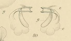
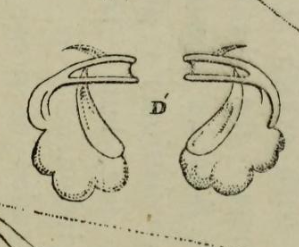
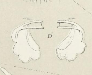
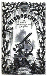
Both Henry James Slack’s Marvels of Pond-Life (1861) and Mary Ward’s Microscope Teachings (1866) copied one of Gosse’s illustrations. They included a drawing of the jaw of a rotifer, a microscopic freshwater animal, which Gosse had made in 1862. Marvels of Pond-Life (1861) introduced beginners in microscopy to freshwater plants and animals, with each chapter focusing on one month of the year when the reader was likely to find the specimen described. Most of the illustrations in the book were made by Charlotte Mary Slack, Henry James Slack’s wife, who combined Gosse’s illustration with drawings of her own. The resulting plate later reappeared in Ward’s Microscope Teachings (1866).

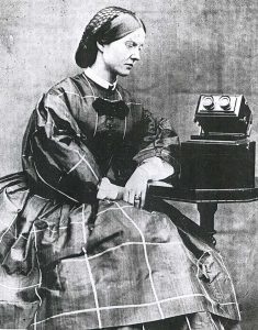
Mary Ward (1827-1869) was born into an Anglo-Irish aristocratic family. She did not receive a formal scientific education, but she dedicated much of her time to making microscopic observations, describing and illustrating them. As a young girl, she occasionally bound her descriptions and illustrations of microscopic specimens into little books and passed them on to friends and family. Later, she revised and published some of her works on microscopy. Microscope Teachings (1866) was one of her most successful publications.

Mary Ward (1827-1869)

Philip Henry Gosse had stressed the importance of field trips for the microscopist, instructing his readers to procure specimens from local ponds or the sea shore. Mary Ward, however, emphasised the domestic side of microscopy, showing her readers how to repurpose things at hand to make microscopic observations. She explained that she “removed the microscope tube from the stand, and mounted it . . . upon a cushion raised on a large book, so that [she] could look as through a telescope into the wine-glass” where she kept microscopic animals. Thus, Ward’s book not only copied Gosse’s illustration but framed it quite differently, as resulting from observations made with the help of household items.

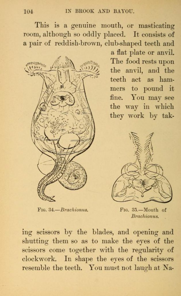
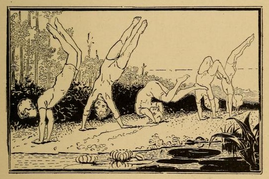
Meanwhile, the illustrations published in Gosse’s popular Evenings at the Microscope (1859) travelled to the United States, where they reappeared in a children’s book, Clara Kern Bayliss’s In Brook and Bayou (1897). Bayliss (1848-1948) was co-editor of the progressive journal The Child-Study Monthly, which advocated for child-centred pedagogy. She edited a section of the journal called the Educational Current, where she commented on developments in child education. Bayliss wrote several children’s books that often blurred the line between fact and fiction and gave ample room to her young readers’ imagination.

Illustration of the microscopic boy in Clara Kern Bayliss’ In Brook and Bayou (1897)

As Bayliss explained in the Educational Current of May, 1899, she regarded a child’s made-up stories as “fiction in its earliest and crudest form, poems and novels by untrained hands”. Instead of teaching children how and what to see through the microscope, Bayliss guided them through a microcosm inhabited by minute animals, mermaids, and a boy shrunk to microscopic size, who was himself microscopically examined by microorganisms. Thus, in Bayliss’s book, Gosse’s illustrations no longer testified to the author’s ability to make truthful scientific observations. Instead, they became an invitation to explore a spectacular, often fictive, microscopic world and to train the imagination rather than the eye.

“A Visit to the Shops”: James W. Queen & Co.

On entering the shop of James W. Queen & Co. at 924 Chestnut Street, Philadelphia, a customer in need of a microscope may have felt overwhelmed by their choices. Founded in 1853, James W. Queen & Co., retailer and manufacturer of scientific instruments of all kinds, stocked simple microscopes (little more than magnifying glasses), compound microscopes, including monocular and binocular instruments, and microscope accessories made by American, English, French and German manufacturers. Customers could choose among watchmaker’s glasses, simple flower microscopes, linen provers (“for counting the threads in linen fabrics”), school and student’s microscopes, dissecting microscopes, portable travelling microscopes, family microscopes and clinical microscopes.

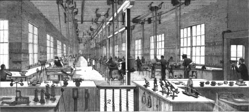
The instrument of the customer’s choice could then be fitted with technical accessories ranging from turn-tables for making specimen slides to watered dissecting troughs and polarising prisms for mineralogical analyses. In 1888, the Scientific American praised James W. Queen & Co. as the “largest and most comprehensive [business] of its kind in the United States or the world.” Their production of scientific instruments had “reached proportions which can hardly be appreciated without a visit to the shops.” Still, the Scientific American tried to recreate the experience of visiting James W. Queen & Co. by walking its readers through the different steps of making scientific instruments. The front page of the journal was covered with illustrations of the various departments at James W. Queen & Co.: The company had its own brass foundry, machinery for grinding lenses and spacious, bright rooms for its designers.

Illustration of James W. Queen & Co.’s factory published in the Scientific American (1888). Image from HathiTrust.

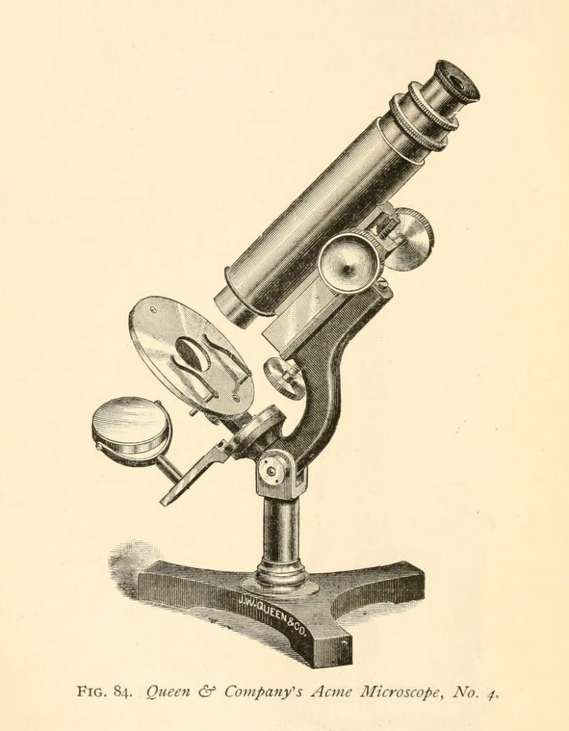
It is unclear if James W. Queen & Co. produced most (or some) parts of their microscopes themselves or if they merely reassembled pieces of instruments produced by other manufacturers. Whatever James W. Queen & Co. did, they did it very successfully. One of the best customers of the company was the US government. The US Army Medical Museum, the Bureau of Animal Industry and the Department of Agriculture were just some of the US departments that relied on microscopes for their daily work. When the article on James W. Queen & Co. was published in the Scientific American, the government had just ordered several Acme microscopes, a series of inexpensive instruments produced by James W. Queen & Co. since 1881. Two years later, Thomas Taylor, official microscopist of the Department of Agriculture, used his Acme microscope to exhibit tea leaves and an embryo tapeworm at the Annual Soirée of the Washington Microscopical Society. Microscopy soirées like this – exhibitions of the latest microscopes, specimen slides carefully prepared for the occasion, or stunning photomicrographs – were often more entertaining than strictly educational. Apparently, governmental microscopical work could also be entertaining. The soirée drew 500 visitors, many of whom “called forth exclamations of delight and wonder.”

Illustration of James W. Queen & Co.’s Acme Microscope No. 4 published in Simon Henry Gage’s (1899) The Microscope and Microscopical Methods. Image from the Internet Archive.

While making microscopes was a difficult task in itself, advertising them was another hurdle microscope manufacturers faced. In addition to circulating regular trade catalogues listing products and prices, James W. Queen & Co. issued a publication that was half trade catalogue and half scientific journal. The Microscopical Bulletin and Optician’s Circular, later The Microscopical Bulletin and Science News, was published from 1883 to 1902 and ingeniously combined price lists, adverts and microscopical news from the US and abroad. Moreover, the wry comments occasionally inserted by the editor, Edward Pennock, made the bulletin a remarkably good read for a trade catalogue turned scientific journal. In 1882, a correspondent of the bulletin enquired: “I want to know if you have a glass that I can see through paper or leather, and if you have one, please to be kind enough to send me the price of it at once.” “Punch a hole in the paper or leather,” Pennock replied.

Since the bulletin was digitised by Google and is not included in the Biodiversity Heritage Library, it will not be uploaded to Worlds of Wonder. Still, it is a delight to read and you can do so in the HathiTrust library.


Further reading

Padgitt, D. L. (1975). A Short History of the Early American Microscopes. London and Chicago: Microscope Publications Ltd. http://www.mccroneinstitute.org/uploads/A_Short_History_Early_American_Microscopes.pdf

Getting to Know the Books and Periodicals on Worlds of Wonder

Philip Henry Gosse: Evenings at the Microscope (1859)

Philip Henry Gosse (1810-1888)

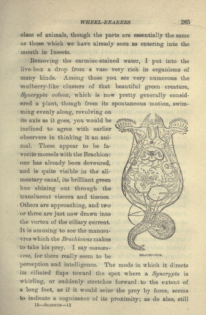
Philip Henry Gosse (1810-1888) was an English zoologist and a prolific author of popular science books. In the early 1850s, Gosse set out to write a book on rotifers, microscopic animals that he collected in ponds and puddles. However, “it proved difficult to popularize so abstruse a subject [as rotifers]” and Gosse abandoned the project. 1 He went on to publish Evenings at the Microscope in 1859, which contains a chapter on rotifers, or “wheel-bearers.” His book was widely read and made rotifer research more popular. In 1897, the illustrations Gosse made for Evenings at the Microscope were reprinted in an American children’s book, Clara Kern Bayliss’ In Brook and Bayou.

Mary Somerville: On Molecular and Microscopic Science (1869)

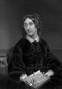
Mary Somerville (1780-1872)

Mary Somerville (1780-1872) was a renowned Scottish astronomer and mathematician, and a celebrated science author. Her On Molecular and Microscopic Science, however, was not as well-received as her books on astronomy and physics. The Popular Science Review called it “a mistake,” its arguments being “crude” and “undigested.” Still, the reviewer admitted that the book’s illustrations were of “great beauty, attractiveness, and effect.” 2 Mary Somerville borrowed heavily from other microscopy handbooks, which makes her On Molecular and Microscopic Science an interesting case of texts and images moving among publications, and possibly being adapted to different audiences.

Thomas Bolton: Hints on the Preparation of Living Objects and Their Examination Under the Microscope (1879-1882)

Thomas Bolton (ca. 1830-1887) was a Birmingham microscopist, who sent living microscopic specimens to microscopists all over Europe. His specimens were accompanied by “flyleaves,” brief descriptions and illustrations of the specimens, often copied from microscopy journals and handbooks. Bolton’s flyleaves were bound and published at intervals. We uploaded a collection of flyleaves circulated between 1879 and 1882. After Bolton died in 1887, his son seems to have taken over his business and continued to send specimens to Europe and North America: There is an advert for “Thomas E. Bolton’s Living Microscopic Specimens” in the American Monthly Microscopical Journal (1898).

The American Monthly Microscopical Journal (1880-1902)

The American Monthly Microscopical Journal was founded by Romyn Hitchcock (1851-1923), a many-sided author, scientist, museum curator and photographer. Originally, the American Monthly Microscopical Journal was associated with the New York Microscopical Society, but in 1887 it was moved to Washington. It contained reports on the meetings of many American microscopy societies, correspondence of its subscribers, descriptions of microscopes and manuals on how to prepare and observe microscopic specimens.

Quarterly Journal of Microscopical Science (1853-1966)

When the Quarterly Journal of Microscopical Science was founded in 1853, it contained the transactions of the Microscopical Society of London and combined them with reports on microscopy in Britain and abroad. However, many members of the London society felt that they should have their own journal and when the society obtained its royal charter in 1866, the quarterly was dissociated from the new Royal Microscopical Society (RMS). The journal continued to be widely read and its successor, the Journal of Cell Science, still exists today.

A “World of Wonder and Beauty”: Exploring Nineteenth-Century Microworlds

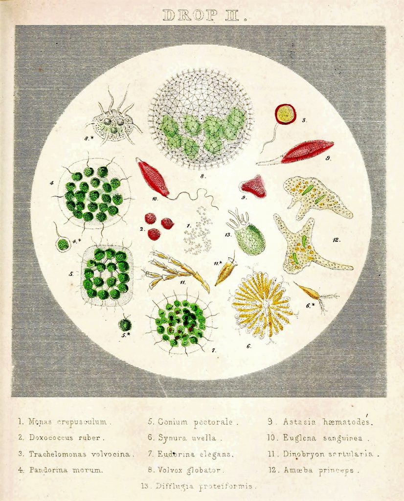
Agnes Catlow (1851) “Drops of water; their marvellous and beautiful inhabitants displayed by the microscope”

In his popular book Evenings at the Microscope (1851), the British microscopist Philip Henry Gosse (1810-1888) described the microscope as “the key that unlocks a world of wonder and beauty” and promised to lead his readers down “the path to the myriad wonders of creation, which . . . are made cognisable to sight by the aid of the Microscope.” 1 Like most nineteenth-century microscopy handbooks, Gosse’s work was richly illustrated. Gosse’s father had been a miniature painter and Gosse himself was a gifted illustrator. His illustrations accompanied several articles he wrote for the Popular Science Review and they featured in his major work on aquatic microorganisms, The Rotifera (1886/1889). 2

Gosse’s illustrations appeared in a whole range of publications aimed at very different groups of readers. Consequently, his illustrations were interpreted differently, depending on the texts that surrounded them and the people who read them. And although Gosse mostly illustrated his articles and books himself, the illustrations that his nineteenth-century readers saw were the result of a collaboration among several people, including the illustrator, lithographer, printer, editor and publisher of a scientific handbook or journal. The sketches Gosse made for his The Rotifera contain instructions to the lithographer, who transferred Gosse’s illustrations to a stone or metal plate and prepared them for printing. 3

Our Worlds of Wonder project seeks to find out more about the ways in which the various people involved in the making of nineteenth-century microscopy illustrations collaborated. Did most microscopists illustrate their own works? How did the decisions made by the editor or publisher affect their illustrations? Did the illustrations change when they were reproduced in another publication? And when did microphotographs start to appear in microscopy publications? With your help, we hope to be able to answer these questions.

Just like Philip Henry Gosse asked his readers to join him on his journey into a “world of wonder and beauty”, we invite you to explore nineteenth-century microscopy publications with us. At the moment, our Zooniverse project contains five publications to choose from, three handbooks and two journals. In our next blog post, we’ll tell you more about these publications and how some of them managed to travel to places all over Europe and America – stay tuned!


Every Book a Compilation: Tracing Microscopy Illustrations

In 1857, the North American Medico-Chirurgical Review published a critique of a microscopy handbook written by the British physician and microscopist Jabez Hogg. The journal criticised Hogg’s publication for copying entire paragraphs “verbatim et literatim” from an American microscopy manual, Joseph Wythe’s The Microscopist (1851). Although the anonymous reviewer admitted that every microscopy handbook was, to some extent, a compilation of previous works, Hogg was attacked for plagiarising Wythe’s book without even mentioning it as his main source – “a Hogg-ish proceeding certainly.”1

The North American Medico-Chirurgical Review was not the only journal to voice such criticism. Reviewers of microscopy handbooks often exposed and condemned plagiarism. Since international copyright laws were only enforced after the signing of the Berne Convention in 1886 and the passing of the American Chace Act in 1891, the mid-nineteenth-century periodical press and book trade thrived on reprinting material. And some editors publicly defended copy-and-paste practices, arguing that carefully selecting and recombining articles was the true art of editing. Edward Pennock, editor of the Philadelphian Microscopical Bulletin and Science News, outright declared that he could “write much better articles with the scissors than with the pen.”2

As a result, microscopy texts and illustrations travelled between books, periodicals and trade catalogues. Illustrations published in a religious work on microscopy, testifying to the beauty of even the most minute of God’s creations, were reprinted alongside medical illustrations. Likewise, an extract from a textbook for students of biology could appear next to a lively account of infusoria living in a microscopist’s garden pond. Often, microscopy texts and illustrations were adapted to target different audiences. Editors shortened texts, added chapters or prefaces, had photos reproduced as engravings or multiple illustrations combined into one. By the time the North American Medico-Chirurgical Review published their scathing review of Hogg’s microscopy manual, the frontispiece of Hogg’s book had already reappeared in a German publication.


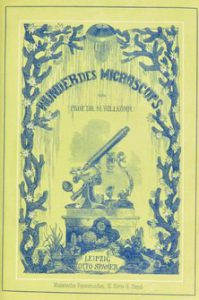
Jabez Hogg’s The Microscope (1854) and Moritz Willkomm’s Die Wunder des Mikroskops (1856)

The Worlds of Wonder Zooniverse project aims to reconstruct the travels of nineteenth-century microscopy illustrations by tracking their reproductions. It builds on the idea that circulating illustrations were a vital factor in the formation of a microscopy community in the nineteenth century. Microscopists were an exceptionally diverse group ranging from amateur and professional scientists in various disciplines to opticians, engineers and medical practitioners. There is reason to believe that the success of microscopy publications depended on their ability to address their readership by combining texts and images which, on their own, would have been relevant only for subgroups within the microscopy community. By reconstructing the routes microscopy illustrations took and documenting the changes they underwent on their way, we hope to better understand strategies of community-building in nineteenth-century science, technology and medicine.

We invite you to join our research team on Zooniverse and help identify and classify microscopy illustrations. Identifying the content and contributors of these illustrations will allow us to get a better idea of who was involved in the nineteenth-century microscopy print trade and to find and analyse reproductions of illustrations. We look forward to meeting you all and having a great time researching microscopy books, periodicals and catalogues together!